


QUICK LINKS
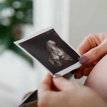
What is an OB Ultrasound?
An obstetric ultrasound, also called sonography, is a type of medical ultrasound and is a medical test that produces pictures of a pregnant woman’s baby, uterus, ovaries, and other structures. This ultrasound examination is usually performed for all pregnant women at 20 weeks gestation, which is typically in the second trimester, to check that your baby is growing properly in your uterus. Obstetrical ultrasound imaging is a key tool for prenatal diagnosis, including the assessment of gestational age and fetal development. It is also performed for many other reasons, including checking the baby’s position in the womb, identifying the number of babies in the womb, first-trimester screening for chromosomal abnormalities, determining the gender of the baby, monitoring the fetal heart and blood flow—often utilizing doppler sonography—and more. Most ultrasound exams are safe, noninvasive, and commonly performed during pregnancy. The use of ultrasound can also provide three-dimensional images in some cases. A doctor trained in radiology will interpret radiology exams, including obstetric radiology exams, to provide accurate results. This procedure is completely painless and non-surgical and is an important part of monitoring your pregnancy.
“I highly recommend Dr. Tepper!He is highly professional, experienced, and calming. My pregnancy and birth were such a positive experience thanks to him and his lovely staff at the clinic.”
SEE MORETypes of Ultrasound Procedures
Ultrasound technology offers a variety of procedures designed to provide detailed images of the body’s internal organs and soft tissues. Each type of ultrasound exam uses high-frequency sound waves to create real-time pictures, helping doctors diagnose medical conditions, monitor pregnancy, and guide certain procedures—all without the use of ionizing radiation. Here’s a look at some of the most common ultrasound procedures:
- Obstetric Ultrasound: Specifically designed for pregnant women, obstetric ultrasound (also called prenatal ultrasound) uses sound waves to monitor fetal growth, check for birth defects, and assess the health of the uterus and amniotic fluid. This type of ultrasound imaging is essential for tracking the development of the fetus throughout pregnancy.
- Transvaginal Ultrasound: For a closer look at the pelvic organs during early pregnancy or when more detailed images are needed, a transvaginal ultrasound may be performed. This procedure uses a special ultrasound probe inserted into the vagina, allowing for clearer pictures of the uterus, ovaries, and other pelvic structures.
- Prenatal Ultrasound: Similar to obstetric ultrasound, prenatal ultrasound focuses on monitoring fetal growth, detecting birth defects, and evaluating the health of the pregnancy. These ultrasound exams are a routine part of prenatal care and provide valuable information about the developing baby.
- Pelvic Ultrasound: Used to assess the uterus, ovaries, bladder, and other pelvic organs, pelvic ultrasound is helpful in diagnosing conditions like ovarian cysts, uterine fibroids, or other abnormalities in the pelvic region.
All of these ultrasound procedures rely on the use of high-frequency sound waves, which bounce off tissues and are converted into electrical signals to create detailed ultrasound images on a computer screen. Because ultrasound exams require no ionizing radiation, they are considered safe for most patients, including pregnant women. Whether you need to evaluate blood flow, monitor fetal growth, or diagnose a medical condition, ultrasound imaging provides clearer pictures and valuable insights for both patients and doctors.
Before Your OB Ultrasound
Before your ultrasound appointment, you may be asked to drink 4-6 glasses of water to fill up your bladder. Having a full bladder can improve image clarity, especially when viewing the urinary tract or reproductive organs. For some ultrasound examinations, you may also be asked to fast for several hours beforehand, depending on the type of scan. Dr. Tepper will provide instructions prior to your appointment specific to your particular case to ensure the best results are achieved. Because an OB ultrasound is an important part of prenatal care, these imaging tests are generally covered under insurance.
The OB Ultrasound Procedure
During your test, you will lie down on a comfortable exam table, and a water based gel will be applied to your abdomen. This gel helps eliminate air pockets between the transducer and your skin, ensuring proper contact for accurate imaging.
A device called a transducer will then be used to send ultrasound waves (high-frequency sound waves) into your body. The transducer sends these sound waves, which bounce off internal tissue and structures, including your baby. The sound waves bounce back to the transducer, and the returning ultrasound signal is processed by the computer to create real-time images for you and your doctor to view.
In most instances, the ultrasound will be performed on the surface of the skin, though a transvaginal ultrasound may be required in some cases. In these instances, a probe may be inserted into a body opening, such as the vaginal canal, to obtain clearer images of the uterus and ovaries. This option may be used early on in pregnancy to determine how far along you are in your pregnancy or to diagnose a suspected problem. Ultrasound also helps visualize and target specific tissue or lesions within the body for diagnostic purposes.
Once the OB ultrasound is completed, the gel will be removed from your skin and you will be able to use the restroom. The test generally takes about 30 minutes to complete.
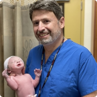
MEET DR. ALEX TEPPER
Welcome to my practice. Let me introduce myself and my practice philosophy. I am a board certified OB/GYN in practice for over 20 years and have been a solo-practitioner on the Upper East Side of Manhattan since 2000. All deliveries are performed at The Mount Sinai Medical Center where I went to medical school and at Lenox Hill Hospital.

When Should I Get an OB Ultrasound?
If you are pregnant and are not sure when you should receive an OB ultrasound, be sure to speak with your OBGYN. In general, women usually undergo ultrasounds following this schedule:
- Early pregnancy: The first ultrasound scan of your pregnancy may be performed during your first trimester, around 6-8 weeks. During this ultrasound scan, your doctor will listen for your baby’s heartbeat, determine gestational age for a more accurate due date, tell you if you are pregnant with multiples, and will determine if an ectopic pregnancy is present.
- Dating ultrasound: If you did not receive an early pregnancy ultrasound, this may be your first ultrasound scan appointment, which is around 10-13 weeks of pregnancy. During this test, Dr. Tepper can estimate gestational age to predict your due date, reveal the number of babies in the womb, check for your baby’s heartbeat, and examine the baby’s basic anatomy.
- Anatomical survey: This OB ultrasound scan is performed between 18-20 weeks. During this test, your doctor will examine the anatomy of your baby, take measurements of the baby, monitor fetal development, and look for physical characteristics that may indicate any abnormalities.
Our Blog

Urinary Tract Infection Treatment

Menopause Management Results: What to Expect

IUD Insertion in Manhattan, NY
Frequently Asked Questions
Do obstetricians do ultrasounds?
How early can fetal abnormalities be detected?
How long does a prenatal ultrasound take?
What is the purpose of prenatal ultrasound?
Can birth defects be seen on ultrasound?
Can having too many ultrasounds hurt the baby?
Is an ultrasound considered routine prenatal care?
Is an ultrasound the same as a sonogram?
How do I prepare for an ultrasound?
What abnormalities can be detected on an ultrasound?
What are signs of abnormal pregnancy?
Learn More About OB Ultrasounds with Dr. Alex Tepper
For more information about what to expect during your OB ultrasound, or to schedule an appointment with Dr. Tepper, contact our New York office today at (212) 828-0900.
BOOK AN APPOINTMENT
Whether you are a new or returning patient, Dr. Tepper can assess your concerns and discuss your options to find the most comfortable and convenient care for you. To get started, call our office to set up an appointment.
CALL FOR APPOINTMENT BILLING & INSURANCE